
Updated September 17, 2026 — Written and reviewed by Dr. Shabbir Hussain, BPT, Registered Physiotherapist | Clinical Rehabilitation Specialist
Quick Answer – Chronaxie vs Rheobase
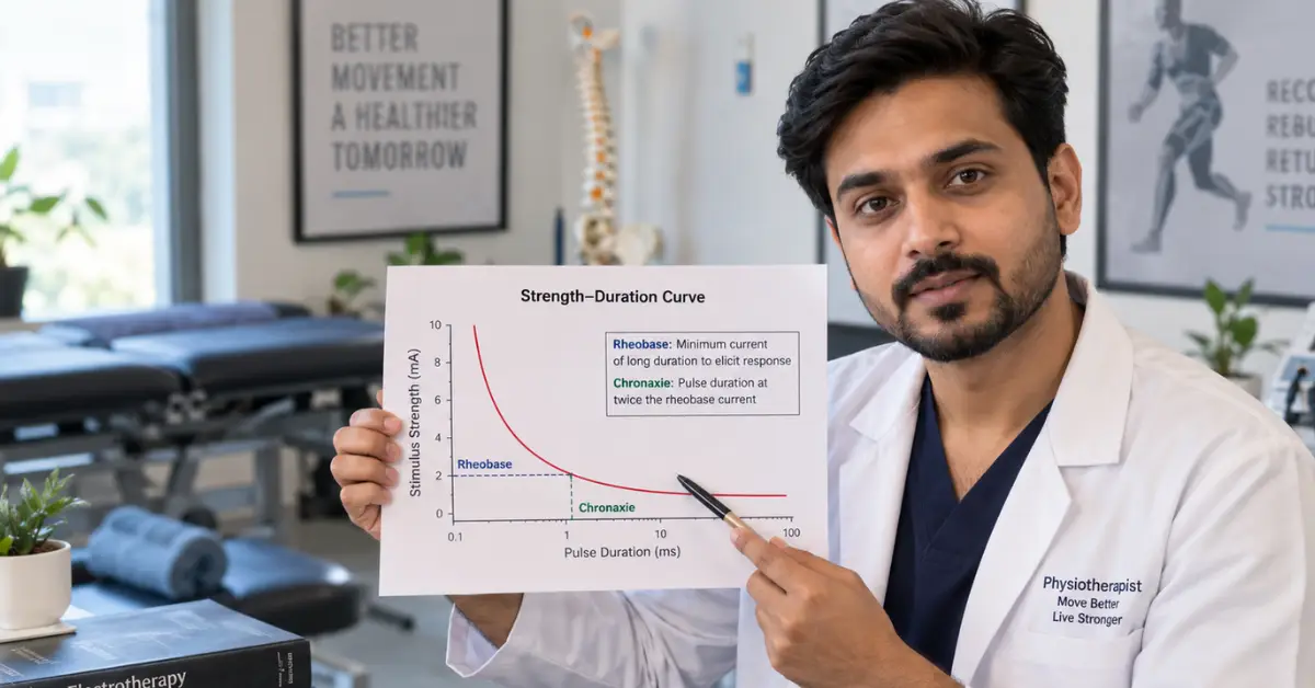
Rheobase is the minimum electrical current strength (at an infinitely long pulse) needed to make a nerve or muscle respond. Chronaxie is the minimum pulse duration needed to get a response when the current is set at exactly twice the rheobase. Together, these two values are plotted on a strength-duration curve, a tool physiotherapists and neurophysiologists use to check how excitable a nerve or muscle is — and, importantly, whether a muscle has lost its nerve supply.
What Is Rheobase?
Rheobase is the threshold. It’s the smallest possible stimulus strength — measured in milliamps or volts depending on the machine — that will still trigger a muscle twitch, no matter how long you leave the current running. Graphically, it’s the point where the strength-duration curve flattens out (asymptotes) along the current axis. Below rheobase, nothing happens, however long the pulse lasts.
What Do You Mean by Chronaxie?
Chronaxie answers a different question: given a stimulus set at twice the rheobase strength, how short can the pulse be and still cause a response? That duration — usually a fraction of a millisecond in healthy tissue — is the chronaxie. It’s often described as a measure of tissue excitability: Chronaxie is one measure of tissue excitability; changes in chronaxie can occur with altered innervation, but the value should not be interpreted in isolation.
A simple way to remember the two: “rheo” relates to current, and “base” to the foundation or minimum strength needed. “Chron” relates to time, and chronaxie is that time value read off the curve at twice-rheobase intensity.
Chronaxie vs Rheobase: The Key Difference
Rheobase is a current (or voltage) value; chronaxie is a time value. Rheobase tells you the minimum intensity a nerve or muscle needs. Chronaxie tells you how quickly it can respond once you double that intensity. Chronaxie varies considerably with the tissue and measurement method.
It is generally shorter in normally innervated excitable tissue and can become substantially prolonged with denervation. When a muscle loses its nerve supply, it becomes a “slower,” less excitable structure electrically — its chronaxie rises sharply, sometimes many times higher than normal, while rheobase typically climbs more gradually.
How to Calculate Chronaxie and Rheobase
Both values come from plotting a strength-duration curve — a graph of the minimum current needed to produce a visible or palpable muscle twitch at a series of different pulse durations.
- Deliver stimuli of varying duration and record the minimum current that produces a twitch at each duration.
- Plot current (y-axis) against duration (x-axis). The curve flattens toward a baseline current value at long durations — that baseline is the rheobase.
- Double the rheobase value.
- Find the pulse duration on the curve that corresponds to this doubled current — that duration is the chronaxie.
Modern electrodiagnostic units can automate this, but the underlying logic hasn’t changed since the concept was first described in early 20th-century neurophysiology.
| Feature | Rheobase | Chronaxie |
|---|---|---|
| Measures | Stimulus strength | Stimulus duration |
| Related to | Minimum threshold intensity | Time needed at 2× rheobase |
| Unit | mA/V depending on method | ms |
| Derived from | Strength-duration curve | Strength-duration curve |
| Clinical meaning | Threshold/excitability information | Time-related excitability information |
| Interpretation | Should be considered with other findings | Should be considered with other findings |
Clinical Applications of Strength-Duration Testing
Strength-duration testing has a real, practical role in physiotherapy and rehabilitation, particularly in peripheral nerve injuries (such as facial nerve palsy, brachial plexus injury, or nerve compression) and spinal cord–related neuromuscular changes.
- Detecting denervation: A rising chronaxie and altered strength-duration curve shape can help identify muscles that have lost some or all of their nerve supply, even before changes are obvious clinically.
- Monitoring recovery: Serial testing over weeks can show a chronaxie value trending back toward normal, which is often an encouraging sign of reinnervation.
- Guiding electrical stimulation: Denervated muscle needs much longer pulse durations to respond than healthy, nerve-intact muscle. Chronaxie values help a physiotherapist select appropriate pulse widths for neuromuscular electrical stimulation (NMES) rather than using a one-size-fits-all setting.
- Who benefits: Patients recovering from peripheral nerve injury, facial palsy, or nerve-related weakness where electrotherapy is part of the rehabilitation plan.
- When to seek assessment: Any new, persistent muscle weakness, drooping, or loss of movement following an injury or illness should be evaluated by a qualified professional before starting any electrical stimulation program — the right settings depend on an accurate diagnosis, not guesswork.
Strength-duration testing is a supportive diagnostic and monitoring tool; it doesn’t replace nerve conduction studies, EMG, or a full clinical neurological examination where those are indicated.
Medical Sources
- Geddes LA — “Accuracy limitations of chronaxie values,” IEEE Transactions on Biomedical Engineering
Link: https://pubmed.ncbi.nlm.nih.gov/14723507/ - PMC (National Institutes of Health) — “Electrical stimulation and denervated muscles after spinal cord injury”
Link: https://pmc.ncbi.nlm.nih.gov/articles/PMC7059583/ - Physiopedia — “Strength-Duration Curve”
Link: https://www.physio-pedia.com/Strength-Duration_Curve
Medical Disclaimer
This article is for general educational and informational purposes only. It is not a substitute for professional medical diagnosis, treatment, or individualized medical advice. If you have persistent, severe, worsening, or concerning symptoms, consult a qualified healthcare professional. Seek urgent medical care for emergency symptoms.

Dr. Shabbir Hussain, BPT
Licensed Physiotherapist | Clinical Rehabilitation Specialist
Maharashtra OTPT Council Reg. No.: PR-2021/08/PT/009532
Society of Onco Physiotherapists Reg. No.: SOP/00033/LM
Dr. Shabbir Hussain is a licensed physiotherapist with over 8 years of experience in physiotherapy, kidney rehabilitation, oncological rehabilitation, and lymphedema management. He specializes in balance disorders, pain management, musculoskeletal rehabilitation, strengthening programs, and VR-based rehabilitation.