Written by Dr. Shabbir Hussain, BPT, Registered Physiotherapist | Clinical Rehabilitation Specialist
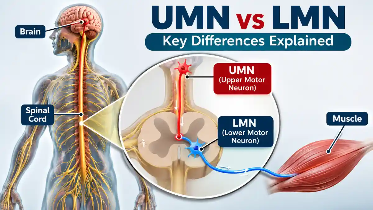
UMN vs LMN – Upper Motor Neurons (UMN) and Lower Motor Neurons (LMN) are two components of the nervous system responsible for controlling voluntary muscle movements. UMN lesions typically cause increased muscle tone, exaggerated deep tendon reflexes and spasticity, whereas LMN lesions cause muscle weakness, reduced muscle tone, diminished reflexes and muscle atrophy.
The key difference is that UMNs control and regulate LMNs, while LMNs directly communicate with skeletal muscles to produce movement.
What Is the Difference Between UMN and LMN?
The difference between UMN and LMN is important in neurological examination because their patterns of muscle weakness, reflexes and muscle tone help clinicians identify where damage has occurred in the nervous system.
Upper Motor Neurons (UMNs) are neurons located in the motor regions of the cerebral cortex and brainstem. Their descending axons travel through pathways such as the corticospinal and corticobulbar tracts to regulate the activity of Lower Motor Neurons involved in voluntary movement.
Lower Motor Neurons (LMNs) are located in the anterior horn of the spinal cord or motor cranial nerve nuclei. Their axons travel through peripheral nerves to directly activate skeletal muscles.
UMN vs LMN: Key Differences
| Clinical feature | UMN lesion | LMN lesion |
|---|---|---|
| Muscle tone | Increased (spasticity) | Decreased (flaccidity) |
| Deep tendon reflexes | Increased | Decreased or absent |
| Muscle weakness | Present | Present |
| Muscle atrophy | Usually mild initially; may develop from disuse | Often prominent |
| Fasciculations | Usually absent | May be present |
| Babinski sign | Usually positive | Usually absent |
| Clonus | May be present | Usually absent |
| Muscle response | Spastic weakness | Flaccid weakness |
| Location | Brain or spinal cord motor pathways above LMNs | Anterior horn cells, motor roots, plexus or peripheral motor nerves |
Important: These are typical clinical patterns, not absolute rules. Acute UMN lesions can initially produce reduced tone and reflexes before spasticity develops. Some neurological conditions also produce mixed findings.
How to Remember UMN and LMN?
A simple way to remember the differences is:
UMN
Upper Motor Neuron = Upregulated reflexes
- Increased tone
- Increased reflexes
- Spasticity
- Positive Babinski sign
LMN
Lower Motor Neuron = Loss of muscle activation
- Low tone
- Reduced reflexes
- Muscle wasting
- Fasciculations
Memory trick: UMN = Hyperactive reflexes; LMN = Hypoactive reflexes.
What Are the Key Differences Between UMN and LMN Palsy?
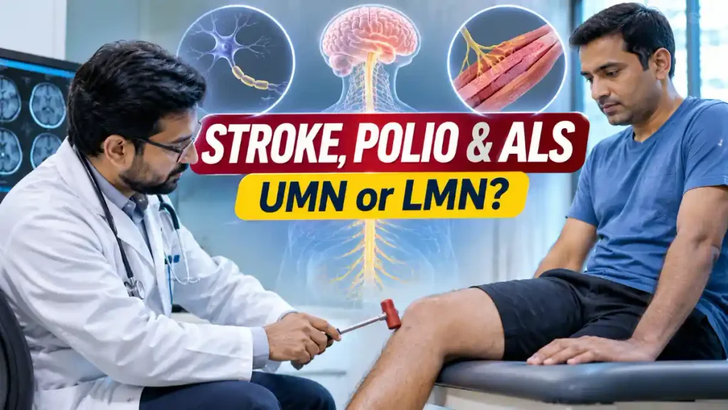
UMN palsy occurs when descending motor pathways in the brain or spinal cord are damaged. LMN palsy occurs when the motor neurons or their axons responsible for directly activating muscles are damaged.
The main clinical differences are:
UMN palsy:
- Spastic paralysis or weakness.
- Increased muscle tone.
- Exaggerated tendon reflexes.
- Positive Babinski sign.
- Clonus may be present.
- Muscle wasting is generally less prominent initially.
LMN palsy:
- Flaccid paralysis or weakness.
- Reduced muscle tone.
- Reduced or absent tendon reflexes.
- Muscle wasting due to denervation.
- Fasciculations may be visible.
- Babinski sign is generally absent in an isolated LMN lesion.
Is Polio an UMN or LMN Disease?
Polio is primarily a Lower Motor Neuron (LMN) disease.
Poliovirus can destroy motor neurons in the anterior horn of the spinal cord and motor nuclei in the brainstem. This results in:
- Acute flaccid paralysis.
- Reduced or absent deep tendon reflexes.
- Muscle weakness.
- Muscle atrophy in affected muscles.
- Usually preserved sensation in paralytic spinal polio.
This explains why paralytic polio produces an LMN-type pattern of weakness.
Is Stroke UMN or LMN?
Stroke typically causes an Upper Motor Neuron (UMN) lesion.
When a stroke damages the motor cortex, internal capsule or corticospinal pathways, it can interrupt signals travelling from the brain to the spinal motor neurons.
Common clinical findings include:
- Weakness or paralysis on the opposite side of the body.
- Increased muscle tone (spasticity).
- Hyperreflexia.
- Positive Babinski sign.
- Difficulty with voluntary and coordinated movements.
However, during the acute stage of stroke, muscle tone and reflexes may initially be reduced. Spasticity can develop later.
The affected side depends on the location of the brain lesion and its relationship to the corticospinal tract crossing in the medulla.
Is ALS Both UMN and LMN?
Yes. Amyotrophic Lateral Sclerosis (ALS) is a motor neuron disease that affects both Upper Motor Neurons and Lower Motor Neurons.
ALS causes progressive degeneration of motor neurons responsible for controlling voluntary muscles.
| UMN features in ALS | LMN features in ALS |
|---|---|
| Spasticity | Muscle atrophy |
| Hyperreflexia | Fasciculations |
| Increased muscle tone | Muscle weakness |
| Positive Babinski sign | Muscle cramps |
| Reduced motor control | Reduced muscle tone in affected muscles |
In ALS, UMN and LMN signs can coexist in the same anatomical region. Reflexes may remain exaggerated even in muscles showing significant wasting because of simultaneous UMN involvement.
Patients may develop progressive weakness, difficulty walking, hand weakness, speech difficulties and swallowing problems.
Importantly, ALS diagnosis requires specialist neurological assessment and appropriate investigations. Having muscle twitching or weakness alone does not establish an ALS diagnosis.
Physiotherapy Rehabilitation in UMN and LMN Disorders
Physiotherapy plays an important role in neurological rehabilitation, particularly for people with UMN and LMN disorders.
For UMN lesions:
- Stretching and positioning to manage muscle tightness.
- Task-specific functional training.
- Balance and gait training.
- Strengthening exercises within the patient’s capacity.
- Spasticity management and appropriate orthotic support.
For LMN lesions:
- Maintaining joint range of motion.
- Carefully prescribed strengthening when appropriate.
- Prevention of contractures.
- Functional mobility training.
- Orthotic support for affected muscles.
Important precautions: Exercise intensity must be individualized. In ALS, excessive fatigue and overwork weakness should be avoided. In acute stroke, rehabilitation should begin under appropriate medical supervision.
Sudden facial drooping, arm weakness, speech difficulty or sudden loss of coordination requires emergency assessment because these can indicate a stroke.
Conclusion
The most important distinction between UMN and LMN lesions is their effect on muscle tone, reflexes and muscle activity. UMN lesions generally produce spasticity and exaggerated reflexes, whereas LMN lesions generally produce flaccidity, reduced reflexes and muscle wasting.
Remember: Stroke = typically UMN, Polio = LMN, and ALS = both UMN and LMN.
MEDICAL SOURCES
1. NCBI Bookshelf — Neuroanatomy, Motor Neuron
Read NCBI reference
2. NCBI Bookshelf — Neuroanatomy, Upper Motor Neuron Signs
Read clinical features of UMN lesions
3. CDC — Poliomyelitis: Epidemiology and Pathogenesis
Read CDC polio reference
4. NCBI Bookshelf — Amyotrophic Lateral Sclerosis Overview
Read ALS reference
5. The ALS Association — Criteria for the Diagnosis of ALS
Read ALS diagnostic criteria
MEDICAL DISCLAIMER
This article is for general educational and informational purposes only. It is not a substitute for professional medical diagnosis, treatment, or individualized medical advice. If you have persistent, severe, worsening, or concerning symptoms, consult a qualified healthcare professional. Seek urgent medical care for emergency symptoms.

Dr. Shabbir Hussain, BPT
Registered Physiotherapist | Clinical Rehabilitation Specialist
Maharashtra OTPT Council Reg. No.: PR-2021/08/PT/009532
Society of Onco Physiotherapists Reg. No.: SOP/00033/LM
Dr. Shabbir Hussain, BPT, Registered Physiotherapist with over 8 years of experience in physiotherapy, kidney rehabilitation, oncological rehabilitation, and lymphedema management. He specializes in balance disorders, pain management, musculoskeletal rehabilitation, strengthening programs, and VR-based rehabilitation.