femur bone anatomy – The femur bone is the longest, strongest, and heaviest bone in the human body.
It is located in the upper part of leg, extending from the hip to the knee .
The femur bone plays a vital role in supporting body weight, enabling movement and protecting various structures.
femur bone anatomy – Main anatomical features :
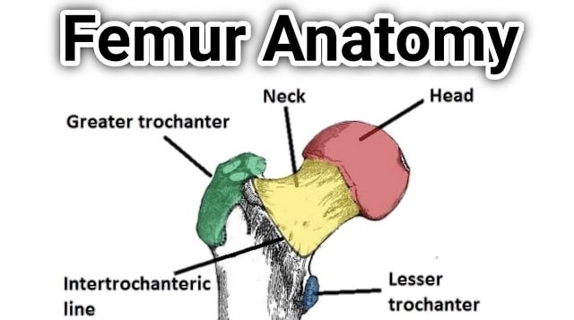
Head:
The rounded, ball-shaped proximal end of the femur bone that forms part of the hip joint.
It articulates with the acetabulum of the hip bone, allowing for a wide range of movements.
Neck:
The narrow section just below the head that connects it to the shaft of the femur bone.
It is angled obliquely, allowing for optimal weight-bearing and transmitting forces from the head to the shaft.
Greater Trochanter:
The bony prominence which is located on the lateral side of the proximal femur.
It serves as an attachment site for various muscles that move the hip and thigh.
Lesser Trochanter:
A smaller bony prominence situated on the posterior and medial aspect of the proximal femur.
Similar to the greater trochanter, it serves as an attachment site for muscles involved in hip and thigh movement.
femur bone Shaft:
The long, cylindrical portion of the femur bone that extends from the neck to the distal end.
It is slightly curved and has a thick cortex to withstand weight-bearing forces.
Medial and Lateral Condyles of femur bone :
The two large, rounded articular surfaces at the distal end of the femur bone.
These condyles articulate with the tibia bone of the lower leg, forming the knee joint.
Intercondylar Notch:
A deep groove located between the medial and lateral condyles at the distal end.
It accommodates ligaments and structures associated with the stability of the knee joint.
Epicondyles:
Bony projections located on the medial and lateral sides of the distal femur, just above the condyles.
These serve as attachment sites for various muscles and ligaments.
Patellar Surface:
The plain smooth, anterior surface of the distal femur which articulates with the patella bone (kneecap).
It forms the patellofemoral joint, allowing for smooth movement of the patella during knee flexion and extension.
Overall, the femur bone is a complex and important bone in the human skeletal system, providing structural support, facilitating movement, and participating in joint articulation.
Important key points in femur Bone viva
When preparing for a viva or an oral examination on the femur bone .
It is essential to understand its key points thoroughly.
Here are some important aspects to consider in viva (femur bone anatomy Simplified ) :
Location and Function:
The femur bone is located in the upper leg and is the longest, strongest, and heaviest bone in the body.
Its primary functions include weight-bearing, supporting the body during movement, and protecting various structures.
femur bone anatomy Simplified :
Familiarize yourself with the different parts of the femur bone, including the head, neck, greater trochanter, lesser trochanter, shaft, medial and lateral condyles, intercondylar notch, epicondyles, and patellar surface.
Articulations:
The femur bone articulates with two main joints—the hip joint and the knee joint.
The head of the femur forms the ball-and-socket joint with the acetabulum of the hip bone, enabling a wide range of movements.
The distal end of the femur articulates with the tibia bone of the lower leg to form the knee joint.
Ligament and Muscle Attachments ( femur bone anatomy Simplified ):
Understand the ligaments and muscles associated with the femur bone.
The greater and lesser trochanters serve as attachment sites for muscles involved in hip and thigh movement.
The epicondyles and intercondylar notch accommodate ligaments and structures associated with knee stability.
Blood Supply:
The femur bone receives its blood supply primarily from the deep femoral artery, which provides oxygen and nutrients to the bone and surrounding tissues.
Fractures and Pathologies:
Be aware of common fractures and pathologies associated with the femur bone.
such as femoral neck fractures, intertrochanteric fractures, femoral shaft fractures and conditions like osteoporosis and osteoarthritis.
Surgical Procedures:
Understand the surgical procedures related to the femur bone, such as total hip replacement, femoral nailing and femoral osteotomy, which are performed to treat fractures, joint diseases, or structural abnormalities.
femur bone Development:
The femur bone develops through a process called endochondral ossification
where a cartilage model is gradually replaced by bone tissue. Understanding the developmental stages can provide insights into growth and potential abnormalities.
femur Bone Clinical Significance :
Familiarize yourself with clinical assessments, imaging techniques like X-rays and MRI scans, and relevant terminology used in diagnosing and treating femur bone-related conditions.
Remember to study these key points thoroughly and ensure a comprehensive understanding of the femur bone anatomy, function and related clinical aspects to excel in your viva or oral examination.
want to know more about Scapula anatomy kindly click here
1 thought on “femur bone anatomy ?”
Comments are closed.