Introduction – ( EMG Explained: How Electromyography Works )
Electromyography, commonly known as EMG, is a valuable diagnostic tool use in the field of medicine, physiology, and biomechanics.
It plays a crucial role in understanding the functioning of muscles and the neuromuscular system.
EMG records the electrical activity generated by muscle contractions, providing insights into muscle health, performance, and various medical conditions.
In this article, we will delve deep into the world of EMG, exploring how it works and its applications.
I. Understanding – EMG Explained: How Electromyography Works
1.1 What is Electromyography?
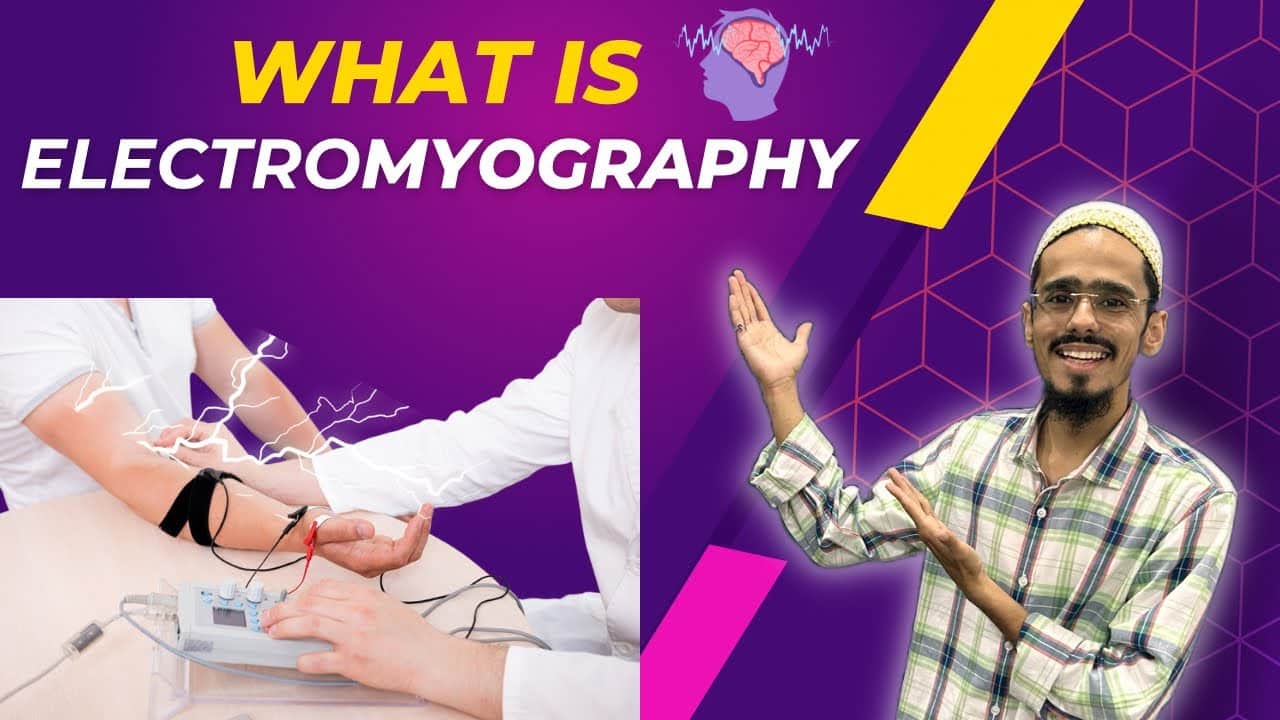
Electromyography, or EMG, is a medical technique use to record the electrical activity of muscles.
The electrical signals, known as electromyograms, provide information about muscle function and activity.
These recordings are essential for diagnosing neuromuscular disorders, understanding muscle function, and assessing muscle performance.
1.2 The Neuromuscular Connection
To comprehend how EMG works, it is essential to understand the neuromuscular system.
Muscles are controll by motor neurons, which are nerve cells responsible for transmitting signals from the brain and spinal cord to the muscles.
When a muscle contracts, motor neurons release electrical impulses, creating a flow of electricity within the muscle fibers. EMG captures and interprets these electrical signals.
II. The EMG Recording Process
2.1 Electrodes
EMG employs electrodes to detect and record muscle activity. There are two main types of electrodes use in EMG:
a) Surface electrodes:
These are place on the skin’s surface and are commonly use for routine EMG examinations.
They are non-invasive but may not provide as detailed information as needle electrodes.
b) Needle electrodes:
These types of electrodes are inserted directly into the muscle tissues.
Needle EMG is more invasive but offers precise data, making it useful for diagnosing specific muscle disorders.
2.2 Signal Amplification and Processing
The electrical signals detected by the electrodes are typically weak.
To make them usable, EMG equipment amplifies and processes the signals.
This step ensures that even the faintest muscle contractions can be record accurately.
2.3 Recording Muscle Activity
During an EMG examination, a trained technician or physician places electrodes on specific muscles of interest.
The patient is then instructed to perform various movements or contractions, such as flexing a limb or maintaining a steady contraction.
As the muscles respond to these actions, the electrodes capture the electrical activity, which will display as a waveform on a computer screen.
III. Interpreting EMG Signals
3.1 Frequency and Amplitude
EMG signals can be analyzed based on their frequency and amplitude.
Frequency refers to the number of electrical impulses produced per second, while amplitude represents the strength or intensity of these impulses.
By examining these characteristics, clinicians can assess the health of the neuromuscular system.
3.2 Motor Unit Recruitment
Muscles are composed of numerous motor units, each controlled by a motor neuron.
EMG helps identify how motor units are recruited during muscle contractions.
This information can reveal abnormalities in muscle function, such as those seen in conditions like muscular dystrophy or motor neuron diseases.
IV. Applications of Electromyography – How Electromyography Works
4.1 Medical Diagnostics
EMG is an indispensable tool in diagnosing a wide range of medical conditions, including:
a) Neuromuscular disorders like amyotrophic lateral sclerosis (ALS).
b) Nerve injuries or compression syndromes.
c) Myopathies, which are diseases affecting muscle tissue.
d) Peripheral neuropathies, where nerves outside the central nervous system are damaged.
4.2 Rehabilitation and Physical Therapy
Physical therapists often use EMG to assess muscle function in patients recovering from injuries or surgeries.
It aids in designing personalized rehabilitation programs and tracking progress.
4.3 Sports Science and Biomechanics
EMG is valuable in sports science and biomechanics for studying muscle activation patterns during different activities.
Athletes can benefit from this data to optimize training regimens and prevent injuries.
V. Advancements in EMG Technology
5.1 Wireless EMG
Recent advancements have led to the development of wireless EMG systems, reducing the need for cumbersome cables and allowing for greater mobility during testing.
5.2 High-Resolution EMG
High-resolution EMG systems offer enhanced sensitivity and precision, making them particularly useful for research and detailed diagnostics.
Conclusion – EMG Explained: How Electromyography Works
Electromyography, or EMG, is a powerful tool that has revolutionized our understanding of muscle function and the neuromuscular system.
By recording and analyzing electrical signals generated by muscle contractions, EMG helps diagnose medical conditions, guides rehabilitation, and advances our knowledge of biomechanics.
With ongoing technological advancements, EMG continues to play a pivotal role in improving healthcare and enhancing our understanding of the human body’s remarkable capabilities.
1 thought on “EMG Explained: How Electromyography Works”
Comments are closed.