Anterior Cruciate Ligament Tear
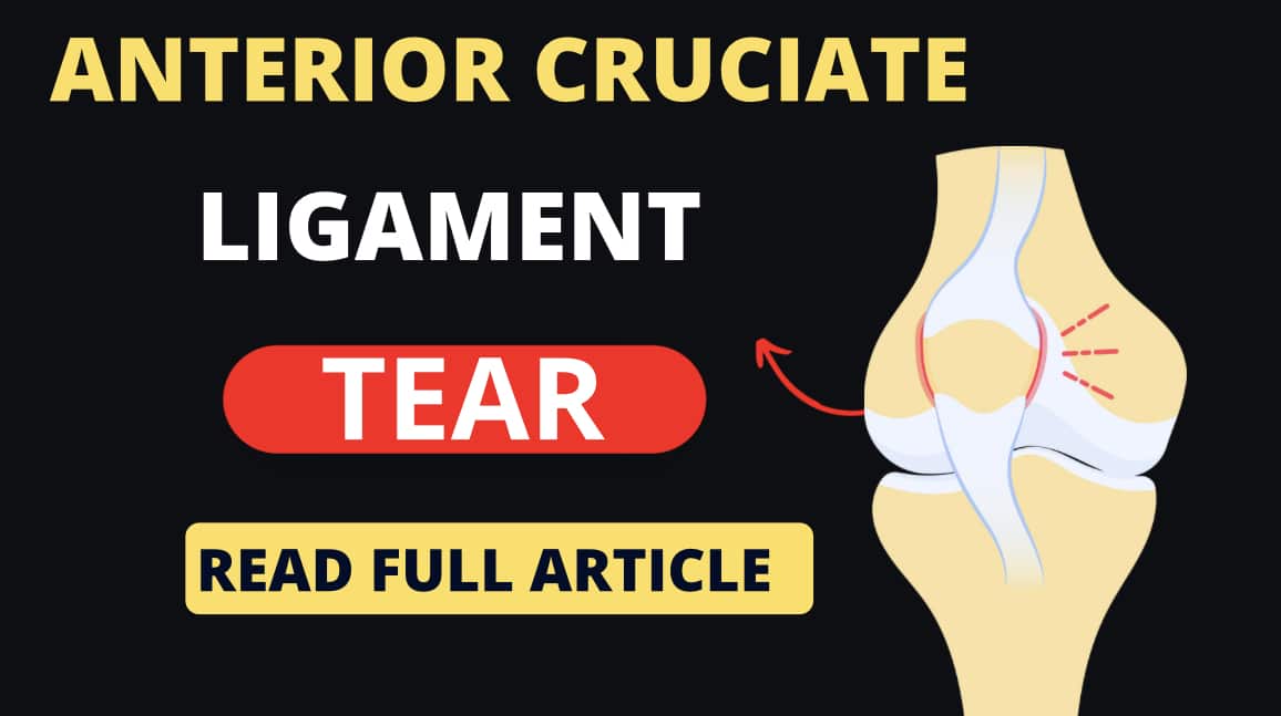
An anterior cruciate ligament (ACL) tear is a common knee injury that affects the ligament connecting the thighbone (femur) to the shinbone (tibia).
The ACL is one of the major ligaments in the knee and plays a crucial role in providing stability to the joint.
Causes:
- Sudden stops or changes in direction while running or jumping
- Direct blow to the knee, such as during a fall or collision
- Landing improperly after a jump
- Twisting or pivoting the knee forcefully
Symptoms:
- A popping and Bursting sound at the time of injury
- Severe pain and swelling in the knee
- Limited range of motion
- Instability or a uneven feeling that the knee may give out
- Difficulty bearing weight on the affected leg
Diagnosis:
A physical examination by a healthcare professional is often the first step in diagnosing an ACL tear.
They will assess the range of motion, stability, and tenderness of the knee. Additional tests may include:
- Imaging tests like X-rays to rule out fractures and MRI scans to evaluate the soft tissues of the knee, including the ligaments.
Treatment:
The treatment for an ACL tear depends on various factors including the severity of the injury, the individual’s activity level, and their overall health. Treatment options include:
- Conservative treatment:
- Protection, Rest, ice, compression, and elevation (PRICE ) therapy to reduce the pain and swelling.
- Physical therapy exercises to restore strength, flexibility, and stability to the knee.
- The use of a knee brace or a supportive device to provide stability during activities.
- Surgical treatment:
- ACL reconstruction surgery is often recommended for individuals who wish to return to sports or have persistent knee instability.
- During the surgery, the torn ligament is replaced with a graft (usually from the patient’s own tissue or a donor) to reconstruct the ACL.
- Rehabilitation and physical therapy are essential after surgery to regain knee strength and stability.
It is important to consult with a healthcare professional, such as an orthopedic surgeon or a sports medicine specialist, Physiotherapist to determine the most appropriate treatment option based on individual circumstances and goals.
Physiotherapy Tests to identify ACL tear
Physiotherapy tests can be helpful in identifying an ACL tear or assessing the stability of the knee joint.
These tests are commonly used as part of the physical examination to evaluate the knee’s integrity and detect possible ligamentous injuries.
Here are a few tests that physiotherapists may perform:
Lachman Test:
The patient lies on their back with the knee flexed at around 20-30 degrees.
The physiotherapist stabilizes the femur with one hand while grasping the tibia below the knee with the other hand.
They then attempt to move the tibia forward, assessing for excessive anterior translation, which may indicate an ACL tear.
Anterior Drawer Test:
The patient lies on their back with the knee flexed at 90 degrees and the foot flat on the table.
The physiotherapist stabilizes the lower leg with one hand while placing the other hand behind the tibia.
They then attempt to pull the tibia forward, assessing for excessive anterior translation compared to the opposite knee.
Pivot Shift Test:
The patient lies on their back with the knee fully extended.
The physiotherapist holds the foot with one hand and places the other hand on the outside of the knee.
They apply a valgus force (inward pressure) to the knee while internally rotating the tibia and slowly flexing the knee.
The test assesses for the presence of a “pivot shift” phenomenon, which is a characteristic giving way or a sudden shift of the tibia that occurs with an ACL tear.
Lateral Pivot Shift Test:
The patient lies on their back with the knee fully extended.
The physiotherapist holds the foot with one hand and places the other hand on the outside of the knee.
They apply a valgus force (inward pressure) to the knee while externally rotating the tibia and slowly flexing the knee.
The test assesses for the presence of a lateral pivot shift, which indicates a combined injury involving the ACL and other structures.
It’s important to note that these tests may provide valuable information, but a definitive diagnosis of an ACL tear often requires further evaluation with imaging studies, such as MRI scans.
A qualified healthcare professional such as a physiotherapist or orthopedic specialist should perform these tests and interpret the results in conjunction with the patient’s history and other clinical findings.
Physiotherapy Protocol for anterior cruciate ligament (ACL) tear
The physiotherapy protocol for an ACL tear typically involves different phases aimed at reducing pain and swelling, restoring range of motion, improving strength and stability and eventually returning to sports or activities.
It is important to note that each individual’s rehabilitation program may vary based on the severity of the injury, surgical intervention and specific goals.
Here is a general outline of the physiotherapy protocol for an ACL tear:
1st Phase : Acute Phase (0-2 weeks)
- Pain and swelling management: RICE (rest, ice, compression, elevation), and possibly the use of anti-inflammatory medication as prescribed by a healthcare professional.
- Promote range of motion (ROM): Gentle active and passive knee exercises to maintain and improve knee mobility, avoiding excessive stress on the ACL.
- Weight-bearing: Gradual progression from partial weight-bearing with the use of crutches to full weight-bearing as tolerated.
2nd Phase: Subacute Phase (2-6 weeks)
- Range of motion exercises: Continue to improve knee ROM, focusing on flexion and extension.
- Strengthening exercises: Initiate gentle quadriceps, hamstring, and calf strengthening exercises to restore muscle function and stability around the knee joint.
- Proprioceptive and balance training: Exercises to improve joint awareness, balance, and coordination.
- Cardiovascular fitness: Low-impact activities, such as stationary cycling or swimming, to maintain cardiovascular fitness without excessive stress on the knee.
3rd Phase: Intermediate Phase (6-12 weeks)
- Progressive strengthening exercises: Gradually increase the intensity and resistance of strengthening exercises, focusing on quadriceps, hamstrings, hip muscles, and core stability.
- Functional training: Incorporate exercises that simulate daily activities and sport-specific movements, gradually adding more dynamic and agility-based exercises.
- Neuromuscular control: Proprioceptive exercises and balance training to enhance control and stability during movement.
- Sports-specific training: Begin sport-specific drills and activities, such as cutting, pivoting, and jumping, under the guidance of a physiotherapist or athletic trainer.
4th Phase : Advanced Phase (12+ weeks)
- Continued strengthening and conditioning: Focus on building strength, power, and endurance specific to the individual’s sport or activity.
- Return to sports/activity: Gradual progression of sport-specific training, including agility drills, running and sport-specific movements.
- Functional testing: Perform various functional tests to assess readiness for return to sports, such as hop tests and agility tests.
- Maintenance program: Develop an ongoing exercise program to maintain strength, stability, and overall fitness to reduce the risk of reinjury.
It’s important to follow the guidance of a qualified physiotherapist or sports medicine professional throughout the rehabilitation process.
They will customize the treatment plan based on individual needs, monitor progress and make adjustments as necessary to optimize recovery and minimize the risk of complications.